
Prostate-specific Membrane Antigen (PSMA) Spatial Biomarker: Predicting Prostate Cancer Survival with Standardized PET Scans
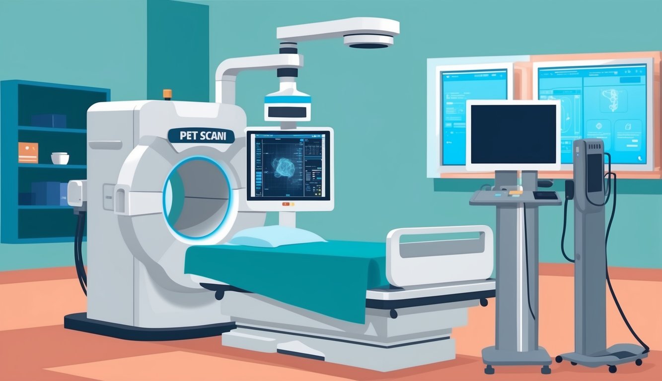
Note: PET scan
A Positron Emission Tomography (PET) scan is a type of imaging test. It uses a radioactive substance called a tracer to look for disease in the body.
A PET scan shows how organs and tissues are working.
This is different than MRI and CT scans. These tests show the structure of, and blood flow to and from organs.
Machines that combine the PET and CT images, called a PET/CT, are commonly used.
Prostate-specific membrane antigen (PSMA) has emerged as a crucial biomarker in the field of prostate cancer research and treatment. PSMA positron emission tomography imaging provides unprecedented accuracy for prostate cancer detection and can offer detailed disease metrics, summarized as a PSMA spatial biomarker (PSMA-SB). This innovative approach to visualizing and quantifying prostate cancer has revolutionized how clinicians assess and manage the disease.
The PSMA spatial biomarker utilizes standardized positron emission tomography disease maps to provide an accurate prediction of overall survival in prostate cancer patients. This advancement allows for more personalized treatment strategies and improved patient outcomes. By incorporating PSMA-SB into clinical practice, healthcare providers can make more informed decisions about treatment options and prognosis.
PSMA PET/CT offers superior diagnostic capabilities for identifying prostate cancer’s spread, with potential as a prognostic indicator for disease recurrence and survival. This technology has transformed the landscape of prostate cancer management, enabling earlier detection and more precise staging of the disease.
Key Takeaways
- PSMA spatial biomarkers provide accurate predictions of overall survival in prostate cancer patients.
- PSMA PET/CT imaging offers superior diagnostic capabilities for prostate cancer detection and staging.
- Standardized PSMA-based disease maps enable personalized treatment strategies and improved patient outcomes.
PSMA in Prostate Cancer: An Overview
Prostate-Specific Membrane Antigen (PSMA) is a protein extensively expressed on prostate cancer cells. It serves as a valuable target for both diagnosis and treatment of prostate cancer.
PSMA expression tends to increase with higher tumor grade and stage. This characteristic makes it particularly useful for detecting advanced and metastatic prostate cancer.
PSMA-ligand PET imaging has revolutionized prostate cancer detection. This technique offers superior accuracy compared to conventional imaging methods, especially for identifying small metastases.
Gallium_PSMA_PET_scan.png
In cases of biochemical recurrence, PSMA-PET can detect tumor sites at lower prostate-specific antigen (PSA) levels than traditional imaging. This allows for earlier intervention and more personalized treatment planning.
For patients with advanced prostate cancer, including castrate-resistant and metastatic forms, PSMA-PET provides crucial information about disease extent and distribution. This data helps guide treatment decisions and assess therapy response.
PSMA’s role extends beyond imaging. It also serves as a target for novel therapies, such as PSMA-targeted radioligand treatment, offering new hope for patients with advanced disease.
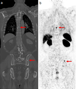
The advent of PSMA-based PET imaging has significantly improved the accuracy of tumor detection. It reveals previously hidden details, including oligo-millimeter nodal and visceral metastases, as well as non-sclerotic bone lesions.
Diagnostics and Staging: PSMA PET/CT’s Role
PSMA PET/CT has revolutionized prostate cancer diagnostics and staging. This advanced imaging technique offers superior accuracy in detecting and localizing prostate cancer lesions compared to conventional methods.
Clinical Applications of PSMA PET/CT
PSMA PET/CT has emerged as a powerful tool for primary staging of prostate cancer. It excels in identifying metastatic disease, including lymph node involvement and distant metastases. The technique’s high sensitivity allows for detection of small lesions that might be missed by conventional imaging.
For patients undergoing radical prostatectomy, PSMA PET/CT aids in preoperative planning. It helps identify extraprostatic extension and seminal vesicle invasion, crucial factors in surgical decision-making.
In cases of biochemical recurrence, PSMA PET/CT proves invaluable for restaging. It can detect recurrent disease at lower PSA levels than traditional imaging methods.
Enhancing Diagnostic Performance
PSMA PET/CT demonstrates superior diagnostic performance compared to conventional imaging techniques. Its high specificity reduces false positives, leading to more accurate diagnoses and treatment plans.
The technique’s sensitivity for detecting lymph node metastases surpasses that of CT or MRI. This improved accuracy in nodal staging can significantly impact treatment decisions.
For local staging, PSMA PET/CT shows promise in determining seminal vesicle invasion and extraprostatic extension. However, its role in this aspect is still being refined and validated.
PSMA PET/CT also enhances the accuracy of prostate biopsies. By identifying areas of high PSMA expression, it can guide targeted biopsies, potentially reducing false negatives.
Overall survival data confirms that PSMA-SB can differentiate aggressive PC from indolent PC
Recent studies have shown that PSMA-based spatial biomarkers (PSMA-SB) derived from PET/CT imaging can effectively distinguish between aggressive and indolent prostate cancer. This differentiation is crucial for tailoring treatment strategies.
Patients with high PSMA expression on PET/CT tend to have more aggressive disease and poorer overall survival. This information allows clinicians to identify high-risk patients who may benefit from more intensive treatment approaches.
Conversely, low PSMA expression may indicate less aggressive disease, potentially allowing for more conservative management strategies. This ability to stratify patients based on disease aggressiveness can help avoid overtreatment in low-risk cases.
The Prognostic Value of PSMA-Based Imaging

PSMA-based imaging has emerged as a powerful tool for predicting outcomes and guiding treatment decisions in prostate cancer. Its ability to detect small metastases and assess tumor burden provides valuable prognostic information.
Predicting Outcomes with PSMA PET/CT
PSMA PET/CT imaging has shown significant promise in predicting overall survival for prostate cancer patients. High PSMA expression on PET scans correlates with poorer prognosis and increased risk of disease progression.
For patients with biochemical recurrence, the number and location of PSMA-avid lesions help determine the risk of treatment failure. Multiple PSMA-positive metastases indicate a higher likelihood of cancer-specific mortality.
PSMA PET/CT also aids in risk stratification. Patients with localized disease but high PSMA uptake may benefit from more aggressive initial therapy. Those with oligometastatic disease on PSMA imaging could be candidates for metastasis-directed treatment.
Treatment Planning and Response Evaluation
PSMA-based imaging plays a crucial role in treatment planning and monitoring response. For radiotherapy, PSMA PET/CT allows for more precise target delineation and dose escalation to PSMA-avid lesions.
In metastatic castration-resistant prostate cancer, PSMA PET can identify patients likely to respond to PSMA-targeted radionuclide therapy. Serial PSMA scans help evaluate treatment efficacy by assessing changes in PSMA expression and tumor burden.
PSMA PET/MRI provides detailed anatomical and functional information for surgical planning. It can detect local recurrence after prostatectomy with high sensitivity, guiding salvage radiotherapy decisions.
PSMA-SB maps could be integrated with multiomics data to improve understanding of total-body cancer heterogeneity and phenotype
PSMA spatial biomarker (PSMA-SB) maps offer a comprehensive view of prostate cancer distribution and heterogeneity throughout the body. Integrating these maps with genomic, transcriptomic, and proteomic data could provide deeper insights into tumor biology.
This multimodal approach may reveal relationships between PSMA expression patterns and specific genetic alterations or molecular subtypes. It could identify distinct phenotypes with different prognoses or treatment sensitivities.
Combining PSMA-SB maps with circulating tumor DNA analysis might enable non-invasive monitoring of clonal evolution and treatment resistance. This integrated approach holds promise for personalized treatment selection and adaptive therapy strategies.
Future Directions in PSMA-Targeted Therapies and Research

PSMA-targeted therapies show promising potential for prostate cancer treatment. Ongoing research focuses on improving theranostic approaches, combining diagnostic imaging and therapeutic interventions.
Molecular imaging techniques continue to evolve, enhancing the accuracy of PSMA-based diagnostics. Researchers aim to develop more sensitive and specific tracers for early detection and monitoring of disease progression.
Clinical trials are exploring novel PSMA-targeted therapies and combination strategies. These include investigating the synergistic effects of PSMA-targeted agents with existing treatments like androgen receptor signaling inhibitors.
Radioligand therapy (RLT) is an area of intense focus. Scientists are working to optimize dosing regimens, reduce side effects, and identify patient populations most likely to benefit from this approach.
Researchers are investigating the potential of PSMA as a biomarker for treatment response and prognosis. This could lead to more personalized treatment strategies based on individual patient profiles.
The interplay between PSMA expression and other molecular pathways, such as DNA repair mechanisms and epigenetic alterations, is being studied. This research may uncover new therapeutic targets and combination strategies.
Efforts are underway to standardize PSMA imaging protocols and interpretation criteria. This will facilitate more consistent results across different centers and improve the clinical utility of PSMA-based diagnostics.
Frequently Asked Questions

PSMA PET imaging has revolutionized prostate cancer management, offering enhanced diagnostic accuracy and treatment planning. This advanced technique provides valuable insights into disease progression and patient prognosis.
How does PSMA PET imaging enhance the management of prostate cancer?
PSMA PET imaging significantly improves the detection and localization of prostate cancer lesions. It offers superior sensitivity and specificity compared to conventional imaging methods.
This enhanced accuracy allows for more precise staging and treatment planning. Clinicians can make better-informed decisions about patient care, potentially leading to improved outcomes.
Can PSMA PET scan predict the overall survival rates for prostate cancer patients?
PSMA PET scans can provide detailed disease metrics that contribute to predicting overall survival in prostate cancer patients. These metrics, known as PSMA spatial biomarkers, offer valuable prognostic information.
The standardized PET disease maps generated from PSMA scans allow for accurate assessment of disease extent and progression. This information aids in estimating survival rates and guiding treatment strategies.
What are the clinical implications of standardized PET disease maps in prostate cancer treatment?
Standardized PET disease maps provide a consistent and reproducible method for assessing prostate cancer spread. They enable more accurate comparisons between patients and across different treatment centers.
These maps help clinicians identify patterns of disease distribution and progression. This information can guide personalized treatment approaches and improve patient management strategies.
How do PSMA expression scores correlate with prostate cancer aggression and prognosis?
PSMA expression scores derived from PET imaging correlate with prostate cancer aggressiveness. Higher PSMA expression is generally associated with more aggressive disease and poorer prognosis.
These scores provide valuable information for risk stratification. They help identify patients who may benefit from more intensive treatment approaches or closer monitoring.
What distinguishes PSMA PET scans from conventional imaging techniques in prostate cancer diagnosis?
PSMA PET scans offer higher sensitivity and specificity compared to conventional imaging techniques like CT and bone scans. They can detect smaller lesions and provide more accurate staging information.
PSMA PET scans also offer whole-body imaging in a single session. This comprehensive approach allows for better assessment of both local and distant disease spread.
In what ways does PSMA-directed therapy influence treatment outcomes for prostate cancer?
PSMA-directed therapy, guided by PET imaging, allows for targeted treatment of prostate cancer lesions. This approach can improve treatment efficacy while minimizing side effects.
It enables more precise radiation therapy planning and can guide surgical interventions. PSMA-directed therapy also opens up new possibilities for targeted drug delivery and radioligand treatments.