
Prostate Cancer – A case study

Disclaimer: Prostate for Dummies is not a medical website. Prostate Cancer – A case study applies to one individual and only relates what he found and what worked for him. This story is told to educate men on prostate cancer and what one individual learned about it. This story should not be taken as medical advice.
The subject was a 52-year-old male who had regular prostate screenings due to yearly physicals that were part of his work insurance program. He was based in Thailand and the physicals were conducted here. Thailand hospitals are very good at data collection and analysis.
In all the years before the discovery of cancer, the subject had no symptoms of any prostate problems, he was healthy and was leading a normal life.
The PSA Test – something every man over 50 should know
PSA refers to Prostatic Specific Antigen. It is part of most physical checkups for men. Average PSA numbers should be investigated. A normal PAS number is between 1 and 4, however, PAS numbers below 1 are the best.
During this checkup, high PSA numbers of ~4 were detected during his yearly physical, but after checking with an ultrasound he did not have an enlarged prostate.

MRI – will that be 1.5 T or 3 T sir?
Note: Because normal PSA numbers are generally between 1 and 4. He was advised to have an MRI to check out the situation.
MRI machines operate with magnetic fields, and radio waves to image the body. The magnetic fields are either a 1.5 T (Tesla) field or a 3 T field. Significant advantages in imaging of prostate cancer can be had with a 3 T machine.
“What does “3T” or “1.5T” refer to?
T (Tesla) refers to the strength of the magnetic field, and higher field strengths improve imaging resolution and the speed of imaging. For imaging of the prostate, field strengths of 3T offer significant advantages in image quality. “
From the University of California, San Francisco Department of Urology website.
The subject was scheduled for an MRI. The MRI included an injection with Gadolinium Dye which helps to pinpoint the location of any prostate cancers, also the cancer can be better characterized due to the contrast produced when using the dye.
Suspected cancerous growth was observed in the subject, he was now officially invested in looking for solutions.
Following the MRI result the subject began researching treatments for prostate cancer. He was told that the cancer was in its early stages and was slow growing. He had some time to research the best procedure for him. He wanted to fully understand what was involved with each treatment before proceeding.
One of the things that the subject found out during his research is that mapping during the MRI process is important to narrowing down and accurately locating the cancer. Detailed mapping is used to pinpoint the cancer and to adjust the biopsy and the treatment.
Once the subject was finished with his research (9 months later) around prostate cancer and treatments he had a full MRI and PSA test done again. This MRI was inconclusive and did not find anything different, however, his PSA numbers had shot up to 9 in the 9 months from 4 when he was first diagnosed.
Tip – Scientists at the Institute of Cancer Research, London (ICR) and the Royal Marsden NHS Foundation Trust have developed a cheap and quick spit test to determine if an individual is susceptible to Prostate Cancer – see the article. This is a very new science. The article was published on June 1st, 2024.
The Apex of the problem
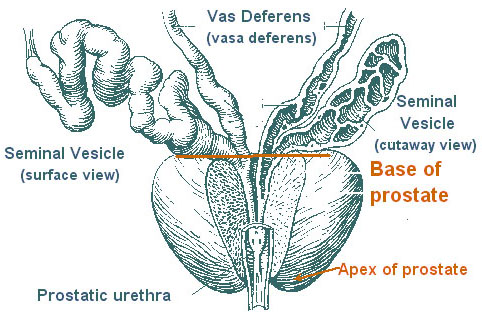
The MRI confirmed that he had lesions on his prostate and needed a biopsy. The biopsy confirmed that the lesion was located at the apex of the prostate gland.
This location is one of the more difficult areas to cure due to the proximity of the sheath of nerves and blood vessels (neurovascular bundles). that surround the prostate. These same bundles play a part in blood flow to the penis and erections.
Yes, that’s right if they make a mistake here, it can have long-lasting complications. If these bundles of nerves are damaged then erections are more difficult to attain, not impossible, just more difficult.
If damage to the neurovascular bundles does occur the problem, can be fixed with medicine but cannot at this time be repaired. Viagra, Cialis, etc.
The MRI shows the lesion (cancer), to be approximately 1.7 x 0.7 cm at the apex of the prostate. A biopsy was performed to determine if the lesion was cancerous.
Biopsy
During a biopsy, samples are taken with a hollow needle in and around the lesion and then randomly around the prostate to see if there are any more areas of interest or any anomalies. This is similar to taking a core of something. A hollow needle is used, and the sample is extracted. This procedure is somewhat unpleasant, however the subject said there was minimal pain.
The subject’s biopsy was comprised of 22 samples. 10 samples were taken in and around the lesion, and 12 samples were randomly spread around the remainder of the prostate. The number of biopsy locations is related to the size of the lesions found and this is one of the reasons that mapping during an MRI is important.
Note: After a biopsy, the prostate will swell up, due to the hollow needle being driven into the prostate in order to take samples of the prostate. It is advised to wait 2 to 3 months after a biopsy before attempting any more treatment or PSA tests to give the time for the swelling to go down.
Our subject had no major complications or discomfort after the biopsy, however, they did piss blood for 4 days (this is normal). He states that he did not feel badly, but did feel slightly uncomfortable.
Gleason Score – Not Jackie Gleeson
The prostate samples are sent to a pathologist to be evaluated for cancerous growth. As with all cancers they are assigned a Gleason Score to determine the severity of the cancer.
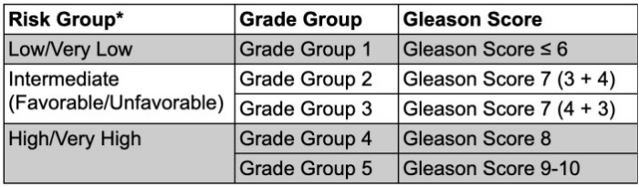
The subject’s Gleason score was 5.
Gleason scores are often a sum of the average of the most affected areas. A Gleason grading determines the aggressiveness of the cancer. The lower the Gleason score the better off you will be.
You will note that the subject was in a low to very low risk group, but, as with many things everything must be taken into account, before making decisions.
Note: A Gleason score higher than 7 normally means that HIFU (the subjects’ choice for treatment may no longer be viable). HIFU is one of the easiest treatments, the least invasive, and the one that provides the best chance for quality of life after treatment. Quality of life refers to still being able to urinate properly, get an erection, and have sex. At 52 the subject was not ready to give up on his sex life just yet.
This is why early detection of prostate cancer is important. If the cancer is too far along HIFU may not be possible.
The subject was understandably concerned regarding any procedure in this area that would probably have a lasting effect on his sex life.
He continued to research the prostate and any procedures available to cure prostate cancer.
Note: His prostate cancer was a slow-growing cancer, so he had time.
Prostate Mapping – Not your Google Map
He decided to send his MRI to several locations to get a second/third opinion of what he had and what was possible.
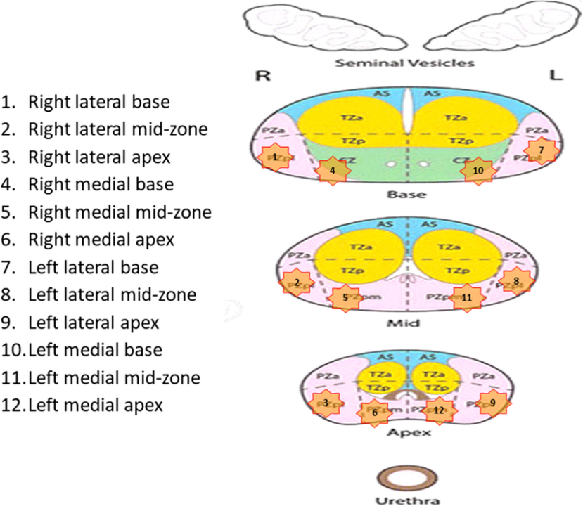
One of the first doctors he spoke to said “Sure send me the data, but be sure to include the mapping data for the MRI”. He was unaware of any mapping data so he went back to the hospital where the MRI was performed and asked them.
It turned out that the mapping had not been done/preserved, and was no longer available. MRI machines keep the data internally for approximately 90 days and the mapping data should have been there, but he missed the deadline and the data was gone.
He requested a second MRI to be performed where the mapping data was made available to him.
A properly performed MRI of the prostate that includes Gadolinium Dye and mapping data can indicate the stage of cancer involved and the precise location. The mapping provides a precise 3-D picture of the prostate.
Note: For anyone who is nervous/claustrophobic the hospital gave the subject a small pill which effectively immobilized him for the entire procedure, with, no ill effects.
Once the redone MRI was submitted to the referral doctor, they confirmed the original diagnosis.
His research continued
Prostate cancer can be treated 3 ways
- Surgery – remove the cancer
- Radiation – target the cancer
- HIFU ultrasound – burn out the cancer
Research, Research, Research
The subject is a meticulous individual and does his research well. In reality, he researched all 3 methods extensively. He had numerous emails and phone calls with doctors about procedures and manufacturers of machines about specifications.
As stated, he spent close to 9 months between work, family, and friends researching all 3 methods and eventually began focusing on HIFU. Due to the location of his cancer, he became aware that HIFU was most likely the best option for a successful outcome in terms of quality of life.
Note: HIFU is not applicable to all people, if the cancer is large and or has moved beyond the prostate gland, then HIFU may not be appropriate. Talk to your doctor.
His research led him to the following conclusions:
- Surgery does work, however, it often results in damage to the neurovascular bundles that surround the prostate (see below). This is not an outcome that you want, and the subject was justifiably concerned. However sometimes surgery is the only option, so be sure to check with your doctors.
- Radiation also does work, however, at present most people are aware that radiation is a bit of a blunt instrument technique. It is possible to target the cancerous area with radiation, but usually, some leakage into the surrounding area happens. Again, the subject was justifiably concerned.
- HIFU (High-Intensity Focused Ultrasound) seemed like the best option for the subject. It is effectively non-surgical and can be performed as an outpatient procedure. The procedure may take from a few to several hours depending on the size of the cancer to be removed. HIFU is not without risks, HIFU ablates (burns) the cancerous cells away. But it can also burn adjacent areas.
All 3 methods have risks associated with them. It is up to every man to assess which method is best suited for them and choose wisely.
See the following article from The Prostate Cancer Foundation.
Interview with an Expert: Rehabilitating your Penis
I encourage you to read the entire article – it is not long – maybe a 5-minute read, and it is not filled with technical mumbo-jumbo.
Note: Surgical techniques (robot), radiation and HIFU are continually evolving, and all of these methods are continually improving. Prostate for Dummies makes no claim that any one method is better than any other. Prostate for Dummies is simply reporting what worked for one man.
Because this article is about one person’s journey with prostate cancer we will focus on the method he chose to remove his cancer HIFU. Both Surgery and Radiation are viable options to remove prostate cancer and there are numerous success stories associated with both methods. HIFU is not applicable to all cases and surgery, or radiation may be your only option. If anything, this article is aimed at letting people know what is available and what works for one person.
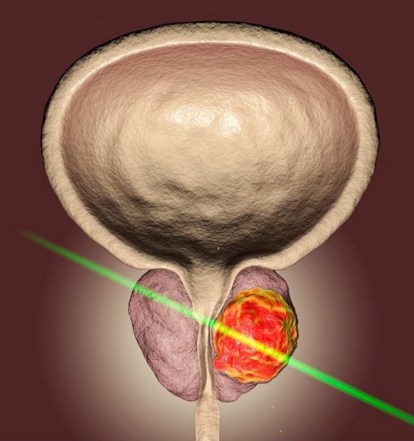
The HIFU Journey – Not Just a Beauty Treatment
HIFU for Beauty
Many people may be aware that HIFU – High-Intensity Focused Ultrasound is commonly used for a beauty treatment. (Just ask your significant other). It is a non-invasive, non-surgical way to provide a facelift. It has been around since 2008 and was approved by the FDA in 2009 for brow lifts. Treatments such as overall facial rejuvenation, lifting, tightening, and body contouring are considered “off-label” (cosmetic), uses and are not yet approved by the FDA.
However, they are commonly done all around the world. The HIFU passes short high-intensity ultrasound waves into the underlying layers of the skin, this heats and causes small amounts of damage to the skin. When the body senses the damage to the skin it produces extra collagen to help with the skin regrowth. The skin tightens up (reducing wrinkles) around that area. The results take a few weeks to achieve the full effect, but this technique has been very successful.
HIFU for Prostate
HIFU for prostate treatment has been approved by the FDA since late 2015. Indeed, HIFU may be covered by your insurance provider. HIFU treatment was covered by insurance for the subject of this article.
HIFU for the prostate works on a similar principle as the facelift HIFU, but the intensity is higher and focused only on the cancerous cells. This allows the cancer to be destroyed, without damaging the surrounding area.
HIFU may apply to localized cancer that has not spread beyond the prostate.
At the time of the subject’s research into HIFU machines, there were 2 main types of HIFU machines – Ablatherm and Sonoblate.
There is a comparison document available here.
Due to the location of the cancer in the subject, his decision was to utilize the Sonoblate machine. It offered a more precise ablation of 0.7 cm vs 1.2cm for the Ablatherm at that time in 2021.
The subject’s research told him several things about the HIFU treatment and the various locations available to him for treatment.
- Thailand – approximately 10 years of experience using the machines.
- Singapore – approximately 10 years of experience using the machines.
- United Kingdom – approximately 20 years of experience using the machines.
The choice seemed obvious – he would travel to the UK for treatment. In his mind, more experience utilizing equipment is always a better choice.
His treatment was at a private clinic in the UK. (that he had researched and found to be the best available). The actual treatment lasted ~2 hours. He felt no ill effects, slightly uncomfortable, but nothing serious. He was able to leave the clinic after approximately 5 hours, which included prep and recovery time. He did have a catheter that needed to be removed after 7 days. He has been well since.
Follow-Up
It is now 2 years after his treatment and his PSA numbers are good (see below). The ongoing success rates for HIFU are quite good for treatment 5 and 10 years after the procedure. Numbers further than that are not yet available. He hopes to be a good statistic for years to come. He continues to send his yearly results to the clinic where he had his procedure done.
He is currently healthy and suffered no lasting ill effects. His PSA number was 1.7, 2 years ago, and is now 1.5 after 2 years.
He plans to actively monitor his prostate with PSA and MRI most likely every year. The long-term plan for my friend is to continue monitoring every 6 months, as suggested with cancer patients. If the cancer returns, he plans to have the surgery again at least one time.
Prostate cancer is a slow cancer, so he feels that he has time. If it does return, he should get a few more years out of another treatment. He will actively monitor his prostate with PSA and MRI most likely every six months or at least every year.
His is a case of early detection leading to a successful result. Not everyone is as lucky as he was.
The news is full of people who did not get tested in time and have died as a result of prostate cancer.
Don’t let yourself be one of them.
Talk to your doctor or your friends. Get the conversation going. Early detection is the key.