Procedures and results explained for patients during An MRI for the Prostate
Prostate MRI is a powerful diagnostic tool that provides detailed images of the prostate gland. This non-invasive procedure uses magnetic fields and radio waves to create high-resolution images, allowing doctors to detect and assess various prostate conditions. Multiparametric MRI (mpMRI) combines four MRI techniques to provide both anatomical pictures and functional information about the prostate gland.
The procedure is primarily used to evaluate prostate cancer, including detection, staging, and monitoring treatment effectiveness. It can also help in identifying other prostate-related issues. Patients undergoing a prostate MRI can expect a relatively comfortable experience, as the procedure is painless and typically takes about 30 to 60 minutes.
MRI is considered the most accurate imaging test for visualizing prostate tissue. It offers superior soft tissue contrast compared to other imaging methods, enabling doctors to make more informed decisions about patient care. This advanced imaging technique has significantly improved the diagnosis and management of prostate conditions, particularly prostate cancer.
Key Takeaways
- Prostate MRI provides detailed images of the prostate gland using magnetic fields and radio waves.
- mpMRI combines 4 MRI techniques for better results.
- The procedure is primarily used for detecting and evaluating prostate cancer, as well as monitoring treatment effectiveness.
- MRI offers superior accuracy in visualizing prostate tissue compared to other imaging methods.
Understanding MRI and Its Role in Prostate Evaluation
Magnetic Resonance Imaging (MRI) has become an essential tool for evaluating prostate health. It offers detailed images of the prostate gland and surrounding tissues, aiding in the detection and characterization of abnormalities.
What Is an MRI?
MRI uses powerful magnets and radio waves to create detailed cross-sectional images of the body. For prostate imaging, patients lie on a table that slides into a large, tube-shaped scanner. The procedure is non-invasive and painless.
During the scan, the magnetic field temporarily aligns the body’s hydrogen atoms. Radio waves then cause these atoms to produce signals, which are used to create detailed images.
MRI scans typically take 30-60 minutes. They provide high-resolution images of soft tissues, making them particularly useful for examining the prostate gland.
The Importance of MRI in Prostate Health
MRI plays a crucial role in prostate cancer management. It aids in:
- Detection and diagnosis
- Guiding biopsies
- Staging of cancer
- Planning treatment
- Monitoring during active surveillance
- Assessing biochemical recurrence
Multiparametric MRI (mpMRI) has emerged as a powerful tool for detecting and localizing clinically significant prostate cancer. It combines multiple MRI techniques to provide a comprehensive evaluation of the prostate.
Definition of an mpMRI:
Here are some details about mpMRI scans:
- What they do mpMRI scans combine four different types of image to create a more detailed picture of the prostate than a standard MRI scan.
- How they work mpMRI scans use a combination of T1-weighted and T2-weighted sequences, diffusion-weighted imaging (DWI), dynamic contrast-enhanced (DCE) imaging, and magnetic resonance spectroscopy (MRS).
- What they show mpMRI scans can show the prostate gland in detail, including the presence of suspicious areas that may be cancer.
- How they’re used Doctors use mpMRI scans to help detect clinically significant prostate cancer, which is the most aggressive type of prostate cancer.
- How they’re scored Doctors give the images of the prostate a score from 1 to 5, called a Likert score. A score of 3 or more usually indicates a biopsy is needed.
MRIs help urologists and radiologists make informed decisions about patient care. It can identify suspicious areas that may require further investigation or biopsy.
MRI vs. Other Imaging Techniques
MRI offers several advantages over other imaging techniques for prostate evaluation:
- Superior soft tissue contrast compared to CT scans
- No radiation exposure, unlike CT scans
- More detailed images than ultrasound
While ultrasound is often used for initial prostate examinations, MRI provides more comprehensive information about prostate structure and potential abnormalities.
Ultrasound alone may miss all or part of a cancerous lesion.
CT scans may be used in conjunction with MRI for staging prostate cancer, particularly to assess lymph nodes and distant metastases.
Recent advancements in MRI technology and interpretation have improved its accuracy in detecting prostate cancer. Artificial intelligence systems are being developed to further enhance MRI interpretation and prostate cancer detection.
The Pre-MRI Process
The pre-MRI process for prostate imaging involves several crucial steps to ensure accurate results and patient safety. These steps include determining the need for the scan, proper preparation, and following essential safety guidelines.
Determining the Need for Prostate MRI
MRI scans are often recommended when there’s suspicion of prostate cancer. Factors that may prompt a doctor to order an MRI include elevated PSA levels and family history of prostate cancer.
PSA levels above 4.0 ng/mL often warrant further investigation. However, the threshold may vary based on age and other risk factors.
Family history plays a significant role. Men with first-degree relatives diagnosed with prostate cancer have a higher risk and may need earlier or more frequent screening.
Preparing for the MRI Scan
Proper preparation is key to obtaining clear, accurate images. Patients should inform their healthcare provider about any medications they’re taking.
Fasting may be required for 4-6 hours before the scan. Patients should avoid caffeine and alcohol for 24 hours prior.
It’s crucial to remove all metal objects, including jewelry, watches, and hearing aids, before entering the MRI room.
Note: If you are claustrophobic, talk to your docotr and ask for something to clam you down before doing the MRI. It will help.
Safety Guidelines and Contraindications
Safety is paramount in MRI procedures. Patients with certain conditions or implants may not be eligible for an MRI.
Metallic implants, such as pacemakers or cochlear implants, can pose risks. Patients should inform their doctor about any implants or medical devices.
Those with kidney problems may need special consideration, as some contrast agents used in MRI can affect kidney function.
Patients with allergies or asthma should disclose this information, as they may be at higher risk for reactions to contrast agents.
Claustrophobia can be an issue for some patients. Sedation options may be available and should be discussed with the healthcare provider.
Inside the MRI Suite
The MRI suite is a specialized environment designed for conducting prostate scans. It houses advanced imaging technology and is set up to ensure patient comfort and safety during the procedure.
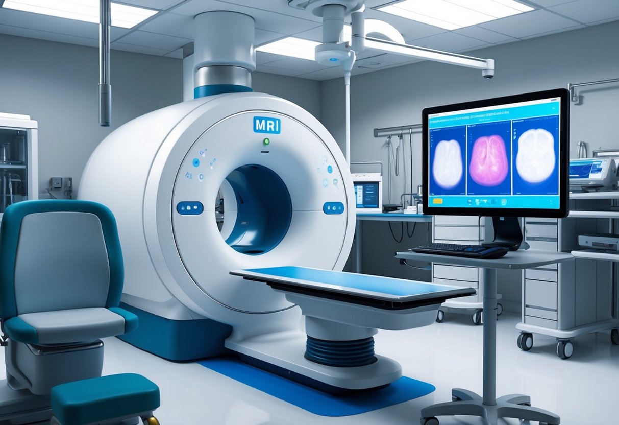
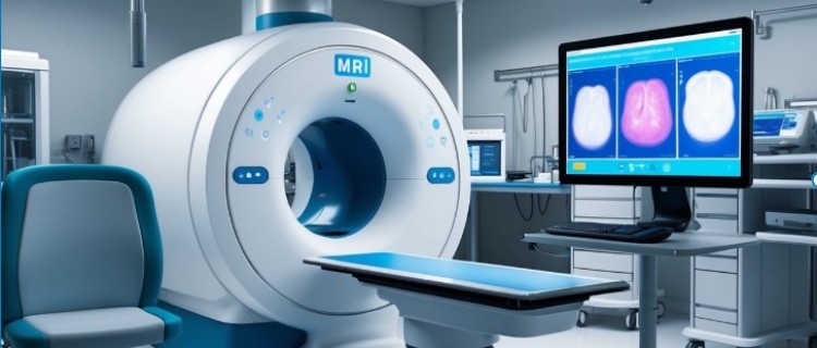
The MRI Equipment
The centerpiece of the MRI suite is the powerful MRI scanner. This large, cylindrical machine uses strong magnetic fields and radio waves to create detailed images of the prostate gland. The scanner includes a movable table where the patient lies during the exam.
Some MRI suites may utilize an endorectal coil, a small device inserted into the rectum to improve image quality. However, many modern scanners can produce high-quality images without this additional equipment.
The control room, separated from the scanning area, houses computer systems for operating the scanner and processing images.
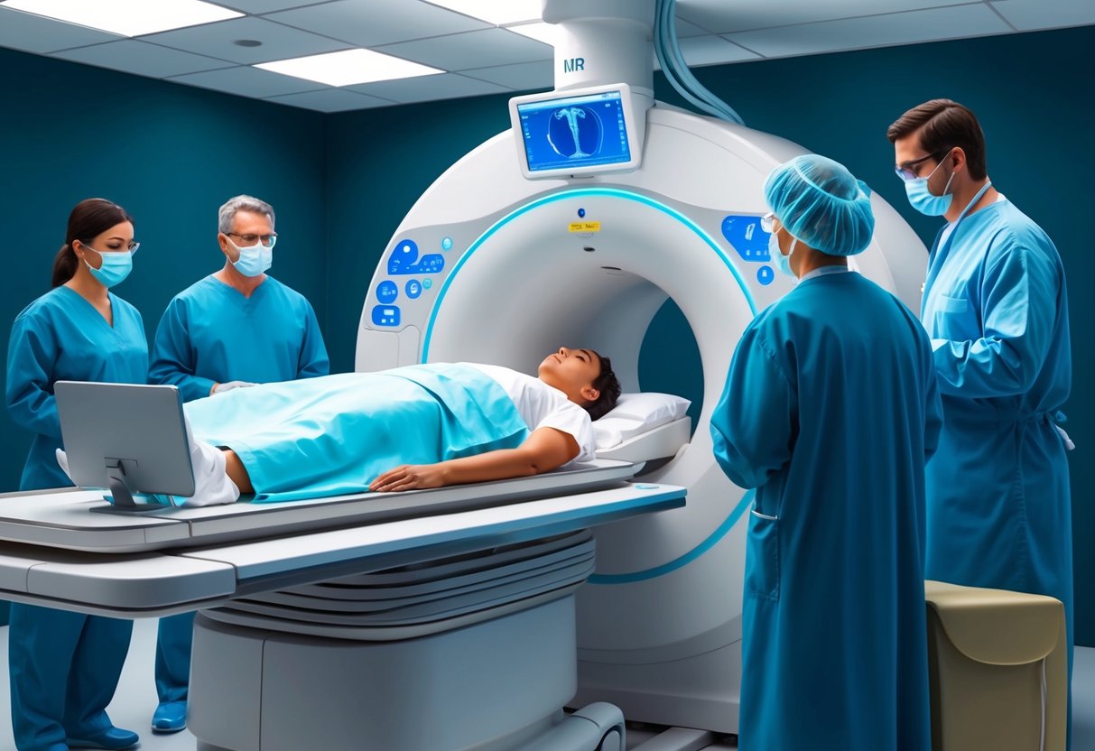
The MRI Procedure
Before entering the scanner, patients change into a hospital gown to eliminate any metal objects that could interfere with the magnetic field. The technologist positions the patient on the table, which then slides into the scanner’s bore.
The MRI technique for prostate imaging typically involves multiple sequences, each capturing different aspects of the gland’s structure and function. These may include T1-weighted, T2-weighted, and diffusion-weighted imaging.
During the scan, the machine produces loud knocking noises as it captures images. Patients wear headphones to protect their hearing and may listen to music to help them relax.
Patient Experience During MRI
An MRI can look and feel scary, especially when you are inside the machine and it is knocking and banging about. Seriously it sounds like the elves are beating each other up in there. The reality is that it is not painful. The hardest part is trying to remain still so that the machine can get a good image of you.
Comfort is a priority during prostate MRI scans, which can last 30-60 minutes. Patients lie still on a padded table, and technologists may provide pillows or cushions for additional support.
Some individuals may experience claustrophobia due to the confined space of the scanner. Techniques to manage this include using wider-bore machines, offering sedation in some cases, or allowing a family member to remain nearby for reassurance.
Communication with the technologist is maintained throughout the procedure via an intercom system. Patients can alert staff if they experience any discomfort or need to pause the scan.
The MRI’s strong magnetic field is not harmful, but it can cause a slight warming sensation or minor tingling in some cases. These effects are normal and temporary.
Comprehensive Prostate MRI Techniques
Prostate MRI techniques have evolved significantly, offering enhanced diagnostic capabilities.
Advanced imaging methods provide detailed anatomical and functional information, aiding in accurate detection and characterization of prostate abnormalities.
Standard MRI vs. Multiparametric MRI
Standard MRI provides basic anatomical information of the prostate gland. However, multiparametric MRI (mpMRI) offers superior diagnostic capabilities by combining multiple imaging sequences.
mpMRI typically includes:
- T1-weighted imaging
- T2-weighted imaging
- Diffusion-weighted imaging (DWI)
- Dynamic contrast-enhanced (DCE) imaging
This comprehensive approach allows for better tissue characterization and improved detection of suspicious lesions. mpMRI has shown higher sensitivity and specificity in identifying clinically significant prostate cancer compared to standard MRI.
Functional Imaging in Prostate Assessment
Functional imaging techniques provide insights into tissue physiology and metabolism. These methods enhance the ability to differentiate between benign and malignant lesions.
Key functional imaging techniques include:
- Diffusion-weighted imaging (DWI): Measures water molecule movement within tissues.
- Dynamic contrast-enhanced (DCE) imaging: Assesses tissue vascularity and perfusion.
- Magnetic resonance spectroscopy (MRS): Analyzes metabolic composition of prostate tissue.
These techniques offer valuable information about tumor aggressiveness and extent, aiding in treatment planning and patient management decisions.
Interpreting MRI Images and PI-RADS
Interpretation of prostate MRI images requires expertise and standardized reporting. The Prostate Imaging Reporting and Data System (PI-RADS) provides a structured framework for evaluating and reporting prostate mpMRI findings.
PI-RADS uses a 5-point scale to assess the likelihood of clinically significant prostate cancer:
- Very low (clinically significant cancer is highly unlikely)
- Low (clinically significant cancer is unlikely)
- Intermediate (the presence of clinically significant cancer is equivocal)
- High (clinically significant cancer is likely)
- Very high (clinically significant cancer is highly likely)
Image quality is crucial for accurate interpretation. Factors such as patient preparation, scanner technology, and imaging protocols significantly impact diagnostic accuracy.
Post-MRI Procedure: Understanding Results and Next Steps

After a prostate MRI, patients enter a crucial phase of result interpretation and decision-making. The process involves skilled professionals analyzing complex images to determine the necessity of further testing or treatment.
Reading and Analyzing MRI Results
Prostate MRI results are typically reported using the PI-RADS scoring system. This 1-5 scale indicates the likelihood of clinically significant prostate cancer. A score of 1 or 2 suggests a low probability, while 4 or 5 indicates a high probability of cancer.
Radiologists also assess tumor location, size, and potential aggressiveness. They use a sector map to pinpoint exact locations within the prostate gland.
Key factors in MRI analysis include:
- T2-weighted imaging
- Diffusion-weighted imaging
- Dynamic contrast-enhanced imaging
These techniques combined provide a comprehensive view of prostate tissue characteristics.
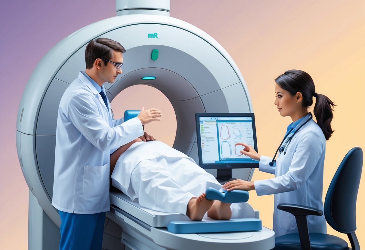
The Role of the Radiologist and Radiographer
Radiologists play a pivotal role in interpreting prostate MRI scans. They analyze images, identify abnormalities, and provide detailed reports to urologists.
High-quality radiology departments with advanced scanners and subspecialty-trained radiologists are crucial for accurate MRI interpretation. Radiographers ensure optimal image quality during the scan.
Radiologists consider factors such as:
- Prostate size and shape
- Presence of lesions
- Extraprostatic extension
- Lymph node involvement
Their expertise helps guide treatment decisions and determine if further investigation is necessary.
When is a Biopsy Recommended?
A biopsy may be recommended based on MRI findings and other clinical factors. Generally, lesions scored PI-RADS 4 or 5 warrant biopsy consideration.
Factors influencing biopsy decisions include:
- PI-RADS score
- PSA levels
- Digital rectal exam results
- Patient age and overall health
MRI-guided biopsies can target specific suspicious areas, improving diagnostic accuracy compared to traditional random biopsies.
For PI-RADS 3 lesions, the decision to biopsy may depend on additional risk factors. PI-RADS 1 or 2 lesions typically do not require immediate biopsy but may be monitored over time.
The Impact of MRI on Prostate Cancer Diagnosis and Treatment
Magnetic resonance imaging (MRI) has revolutionized prostate cancer management. It provides detailed anatomical and functional information, enhancing detection accuracy, guiding treatment decisions, and improving patient outcomes.
MRI in the Detection of Early Prostate Cancer
Multiparametric MRI (mpMRI) has emerged as a powerful tool for detecting early prostate cancer. It combines anatomical and functional imaging sequences to provide a comprehensive assessment of the prostate gland.
mpMRI can identify suspicious lesions that may be missed by conventional methods. This increased sensitivity allows for more targeted biopsies, reducing the need for unnecessary procedures.
The use of mpMRI before biopsy has shown to improve cancer detection rates, particularly for clinically significant tumors. It helps distinguish between aggressive and indolent cancers, aiding in risk stratification.
Local Staging and Treatment Planning
MRI plays a crucial role in local staging of prostate cancer. It provides detailed information about tumor size, location, and extent, which is essential for treatment planning.
Key benefits of MRI in staging include:
- Assessing extracapsular extension
- Evaluating seminal vesicle involvement
- Identifying pelvic lymph node metastases
This information guides treatment decisions, such as choosing between surgery, radiation therapy, or active surveillance. MRI helps determine the feasibility of nerve-sparing prostatectomy and optimizes radiation therapy planning.
Active Surveillance and Recurrent Prostate Cancer
For patients on active surveillance, MRI is increasingly used for monitoring. It helps detect cancer progression early, allowing timely intervention when necessary.
MRI-guided biopsies can confirm the presence of higher-grade tumors, influencing decisions to transition from surveillance to active treatment.
In cases of recurrent prostate cancer, MRI aids in:
- Detecting local recurrence after primary treatment
- Differentiating between local recurrence and post-treatment changes
- Guiding salvage treatments
The high soft-tissue contrast of MRI makes it particularly useful for identifying small areas of recurrent disease, enabling more precise and effective salvage therapies.
Considerations and Advancements in Prostate MRI
Recent developments in prostate MRI have improved image quality and diagnostic accuracy. Innovations in technology and techniques address patient comfort and anxiety during scans. Future advancements promise even more precise and accessible prostate imaging.
New Developments in Prostate Imaging
Multiparametric MRI (mp-MRI) has significantly enhanced prostate cancer diagnosis and staging. This technique combines anatomical and functional imaging sequences to provide detailed information about prostate tissue.
Higher magnetic field strengths, such as 3-Tesla MRI machines, offer improved spatial resolution and signal-to-noise ratios compared to 1.5-Tesla systems. These advancements allow for better detection of small lesions and more accurate characterization of tumor aggressiveness.
Artificial intelligence and machine learning algorithms are being integrated into image analysis. These tools help radiologists identify suspicious areas more efficiently and may improve the consistency of interpretations across different readers.
Dealing with Anxiety and Claustrophobia
MRI scans can be anxiety-inducing for some patients, particularly those with claustrophobia. Modern MRI machines often feature wider bores and shorter tunnels to reduce feelings of confinement.
If you check with your doctor a sedative may be available to help you through the MRI session. It can take a long time to get the results needed, so a sedative may help you to remain still during the procedure.
Strategies to manage anxiety during prostate MRI:
- Providing clear explanations of the procedure
- Offering music or audio entertainment during the scan
- Using breathing techniques or mild sedation when necessary
Some facilities now offer open MRI machines, which can be more comfortable for claustrophobic patients. While these may have limitations in image quality, they provide a viable alternative for those who cannot tolerate traditional closed MRI scanners.
Future Outlook in MRI Technology
Ongoing research aims to further improve prostate MRI technology. Hybrid imaging systems, such as PET-MRI, are being explored to combine metabolic information with high-resolution anatomical images.
Advanced coil designs and faster imaging sequences are in development. These innovations may reduce scan times while maintaining or improving image quality, enhancing patient comfort and increasing throughput.
Molecular imaging techniques using novel contrast agents are under investigation. These may allow for more specific targeting of cancer cells, potentially improving the accuracy of prostate cancer detection and characterization.
Additional Prostate Conditions Evaluated via MRI
Prostate MRI is a versatile diagnostic tool that extends beyond cancer detection. It provides valuable insights into other common prostate conditions, aiding in comprehensive evaluation and management of prostate health.
Benign Prostatic Hyperplasia (BPH) and Prostatitis
Prostate MRI plays a crucial role in assessing benign prostatic hyperplasia (BPH) and prostatitis. BPH, characterized by an enlarged prostate, can be accurately measured and visualized through MRI. The imaging technique allows for precise volume calculations and helps determine the impact on surrounding structures.
For prostatitis, MRI can differentiate between acute and chronic forms. It reveals inflammation patterns, abscesses, and structural changes. This information guides treatment decisions and helps monitor disease progression.
MRI also assists in planning minimally invasive treatments for BPH, such as prostate artery embolization. The detailed anatomical information it provides enhances procedural accuracy and outcomes.
Monitoring PSA and Prostate Health
Prostate-specific antigen (PSA) testing is a common screening tool, but it has limitations. MRI complements PSA monitoring by providing visual context to elevated PSA levels. It can identify non-cancerous causes of PSA elevation, reducing unnecessary biopsies.
Regular MRI scans can track changes in prostate volume and structure over time. This is particularly useful for men on active surveillance or those with a family history of prostate issues.
MRI also aids in evaluating the effectiveness of treatments for various prostate conditions. It can monitor shrinkage in BPH cases or assess the resolution of inflammation in prostatitis.
The combination of PSA testing and MRI imaging offers a comprehensive approach to prostate health management. It allows for more informed decision-making and personalized treatment strategies.
Frequently Asked Questions

Prostate MRI procedures involve specific preparations, techniques, and potential findings. Understanding the process, requirements, and results can help patients feel more informed and at ease.
What is the procedure for conducting a prostate MRI?
A prostate MRI is a non-invasive imaging technique that provides detailed images of the prostate gland. The patient lies on a table that slides into the MRI machine.
The technician may administer a contrast dye intravenously to enhance image quality. The procedure typically lasts 30-60 minutes, during which the patient must remain still.
What types of findings can a prostate MRI reveal?
A prostate MRI can detect and diagnose prostate cancer in its early stages. It can also reveal the size and shape of the prostate gland, identifying any abnormal growths or lesions.
The MRI can determine if cancer is present, its aggressiveness, and whether it has spread beyond the prostate. It also helps in assessing other prostate conditions like benign prostatic hyperplasia.
How should one prepare for a prostate MRI, including dietary restrictions?
Patients should inform their doctor of any allergies, medical conditions, or implanted devices. They may need to avoid eating or drinking for several hours before the exam.
Wearing comfortable, metal-free clothing is advised. Some facilities may provide a gown. Patients should remove all metal objects, including jewelry and watches, before the procedure.
What are the different stages or categories in prostate MRI results?
Prostate MRI results are often categorized using the PI-RADS (Prostate Imaging-Reporting and Data System) scale. This system ranges from 1 to 5, with higher numbers indicating a greater likelihood of clinically significant cancer.
A score of 1 or 2 suggests a low probability of cancer, while 4 or 5 indicates a high probability. A score of 3 is considered intermediate.
What are the reasons for a doctor to recommend an MRI of the prostate?
Doctors may recommend a prostate MRI for early detection and accurate diagnosis of prostate conditions, including cancer. It’s often used when blood tests show elevated PSA levels or after an abnormal digital rectal exam.
MRI can also help in treatment planning, guiding biopsies, and monitoring the progression of known prostate cancer.
What is the typical duration of a prostate MRI exam?
A prostate MRI exam usually takes between 30 to 60 minutes to complete. The exact duration can vary depending on the specific protocol used and whether contrast is administered.
Patients should plan to spend about 90 minutes at the imaging facility to account for preparation time and post-exam procedures.