Transrectal ultrasound (TRUS) is a medical imaging technique used to examine and diagnose conditions in the pelvic area, particularly the prostate gland in men. This non-invasive procedure utilizes high-frequency sound waves to create detailed images of internal organs and tissues.
[Transrectal ultrasound (TRUS) uses high-energy sound waves to create images of organs and tissues, The TRUS procedure involves inserting a small probe into the rectum, which emits sound waves that bounce off nearby structures. These echoes are then converted into real-time images on a monitor, allowing medical professionals to examine the prostate and surrounding areas.
TRUS plays a crucial role in various medical applications, including guiding biopsies, assessing prostate size and structure, and detecting abnormalities such as tumors or cysts. It is often used in conjunction with other diagnostic tools, such as prostate-specific antigen (PSA) tests and digital rectal exams, to provide a comprehensive evaluation of prostate health.
Key Takeaways
- TRUS is a non-invasive imaging technique used to examine the prostate and surrounding tissues.
- The procedure helps diagnose prostate conditions, guide biopsies, and detect abnormalities.
- TRUS is often used alongside other diagnostic tools for comprehensive prostate health evaluation.
Understanding Transrectal Ultrasound
Transrectal ultrasound (TRUS) is a diagnostic imaging procedure that provides detailed views of the prostate gland. It uses high-frequency sound waves to create images, allowing healthcare providers to examine prostate abnormalities and guide biopsies.
The Process and Mechanism of TRUS
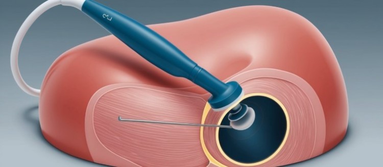
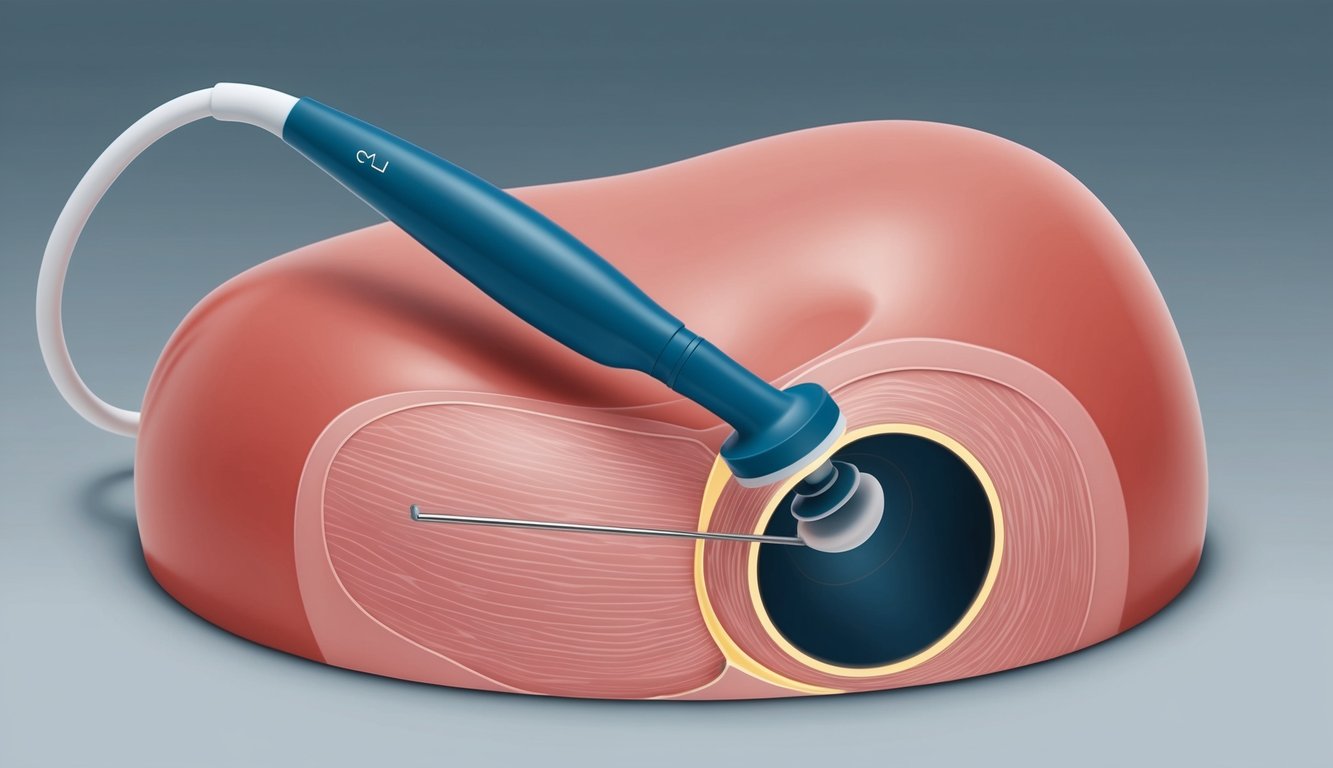
During a TRUS procedure, a small ultrasound transducer is inserted into the rectum. This probe emits sound waves that bounce off the prostate and surrounding tissues. The echoes are then converted into real-time images on a monitor.
The ultrasound waves can penetrate soft tissues, providing clear visualizations of the prostate’s structure and any potential abnormalities. TRUS allows healthcare providers to measure the size of the prostate and identify suspicious areas that may require further investigation.
TRUS is often combined with a Digital Rectal Exam (DRE) for a more comprehensive evaluation. This combination enhances the accuracy of prostate assessments and helps guide needle biopsies if needed.
Comparing TRUS to Other Prostate Imaging Techniques
TRUS offers several advantages over other prostate imaging methods:
- Real-time imaging
- No radiation exposure
- Cost-effective
- Widely available
However, it has limitations compared to advanced techniques like MRI:
- Lower resolution for detecting small lesions
- Less effective in distinguishing between benign and malignant tumors
- Limited field of view
MRI provides more detailed images and can detect smaller abnormalities, but it is more expensive and time-consuming. CT scans use radiation and are less effective for soft tissue imaging of the prostate.
Preparing for a Transrectal Ultrasound
Proper preparation ensures optimal results from a TRUS procedure:
- Inform the healthcare provider of any medications, especially blood thinners.
- Follow instructions regarding food and drink before the exam.
- An enema may be required to clear the rectum.
- Antibiotics might be prescribed to prevent infection, especially if a biopsy is planned.
Patients should wear comfortable, loose-fitting clothing and may be asked to change into a gown. The procedure typically takes 15-30 minutes and is performed on an outpatient basis.
Relaxation techniques can help reduce anxiety and discomfort during the exam. Patients may experience mild pressure or discomfort but rarely pain.
Transrectal Ultrasound in Diagnostics and Treatment
Transrectal ultrasound (TRUS) plays a crucial role in diagnosing and treating prostate conditions. It provides detailed images of the prostate gland, enabling accurate biopsies and guiding various procedures.
TRUS-Guided Procedures and Biopsies
TRUS-guided prostate biopsies are essential for diagnosing prostate cancer. The ultrasound probe guides a needle to collect tissue samples from specific areas of the prostate. Typically, 10 to 18 samples are taken.
Before the procedure, patients may receive local anesthetic to minimize discomfort. Those on blood thinners might need to adjust their medication.
The biopsy process usually takes 15-30 minutes. Patients may experience mild pain or soreness afterward.
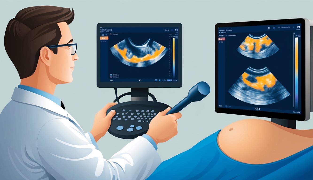
Interpreting TRUS Results
TRUS images can reveal abnormal growths or changes in the prostate gland. Radiologists analyze these images to identify potential cancerous areas.
Biopsy results are typically available within a week. They indicate whether cancer cells are present and, if so, their grade and extent.
TRUS results are often considered alongside other factors, such as prostate-specific antigen (PSA) levels, to determine the best course of action.
Post-TRUS Follow-Up and Care
After a TRUS procedure, patients receive specific follow-up instructions. These may include:
- Avoiding strenuous activities for 24-48 hours
- Taking antibiotics to prevent infection
- Monitoring for side effects like difficulty urinating or blood in urine
Patients should report any severe pain, fever, or excessive bleeding to their healthcare provider immediately.
Follow-up appointments are scheduled to discuss results and plan further treatment if necessary. Regular PSA testing and additional imaging may be recommended for ongoing monitoring.
Frequently Asked Questions
Transrectal ultrasound is a diagnostic procedure used to examine the prostate and surrounding tissues. This imaging technique provides valuable information for detecting and evaluating various prostate conditions.
What is the purpose of a transrectal ultrasound?
A transrectal ultrasound (TRUS) is a key tool for evaluating the prostate gland. It helps healthcare providers visualize the prostate and surrounding tissues.
TRUS can detect abnormalities, guide biopsies, and assist in diagnosing prostate conditions. It is commonly used to investigate prostate cancer, benign prostatic hyperplasia, and prostatitis.
What are the steps involved in a transrectal ultrasound procedure?
The procedure typically takes less than 30 minutes. The patient lies on their side with knees bent towards the chest.
A lubricated probe is gently inserted into the rectum. The probe emits high-frequency sound waves that create images of the prostate on a monitor.
In some cases, a biopsy may be performed during the procedure. The healthcare provider uses the ultrasound images to guide the biopsy needle.
How does a transrectal ultrasound help in diagnosing prostate conditions?
TRUS provides detailed images of the prostate gland’s size, shape, and internal structure. It can reveal abnormal growths, cysts, or areas of inflammation.
The procedure helps detect prostate cancer by identifying suspicious areas that may require biopsy. It also assists in evaluating benign prostatic hyperplasia by measuring prostate volume.
Can transrectal ultrasound be used for diagnostic purposes in females, and if so, what does it detect?
Transrectal ultrasound can be used in females to examine the rectum, anal canal, and surrounding pelvic structures. It may detect rectal tumors, anal fistulas, or abnormalities in nearby organs.
In females, transvaginal ultrasound is more commonly used for examining reproductive organs. Transrectal ultrasound is generally reserved for specific conditions affecting the rectum or anus.
What should one expect in terms of discomfort during a transrectal ultrasound?
Transrectal ultrasounds may cause minimal discomfort, similar to a rectal exam. Patients might feel pressure or a mild sensation when the probe is inserted.
If a biopsy is performed, patients may experience a more intense sensation in the rectum. A numbing agent is typically used to minimize discomfort during biopsies.
What are the potential risks associated with a transrectal ultrasound?
Transrectal ultrasound is generally considered safe with minimal risks. Some patients may experience mild rectal bleeding or discomfort after the procedure.
In rare cases, infection may occur, especially if a biopsy is performed. Proper sterilization techniques and antibiotic prophylaxis help reduce this risk.